Unveiling the Human Body: A Comprehensive Review of the Pocket Atlas of Sectional Anatomy, Volume 1

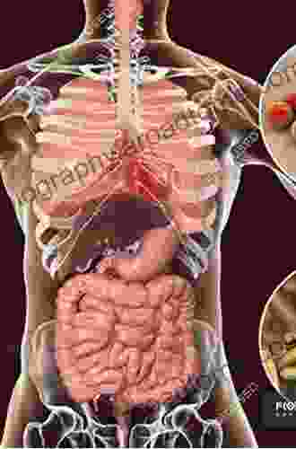
The human body is a marvel of intricate complexity, with countless structures and systems working harmoniously to sustain life. Understanding the anatomy of the human body is essential for medical professionals, students, and anyone interested in the inner workings of our physical form. Sectional anatomy, the study of the body through cross-sectional images, provides a unique perspective that allows us to visualize and comprehend the various anatomical structures in a three-dimensional context.
The Pocket Atlas of Sectional Anatomy, Volume 1, is a comprehensive guide to sectional anatomy, providing an unparalleled depth of knowledge and imagery. This exceptional atlas is a must-have for medical professionals, students, and anyone seeking to expand their understanding of human anatomy.
4.7 out of 5
| Language | : | English |
| File size | : | 82385 KB |
| Text-to-Speech | : | Enabled |
| Screen Reader | : | Supported |
| Enhanced typesetting | : | Enabled |
| Print length | : | 340 pages |
An In-Depth Exploration of Sectional Anatomy
The Pocket Atlas of Sectional Anatomy, Volume 1, encompasses a vast range of cross-sectional images, including:
- Magnetic Resonance Imaging (MRI): MRI utilizes powerful magnets and radio waves to generate high-resolution images of the body's soft tissues and organs.
- Computed Tomography (CT): CT uses X-rays and computer processing to create detailed cross-sectional images of the body's bones and tissues.
- Ultrasound: Ultrasound employs sound waves to produce real-time images of the body's internal structures.
These advanced imaging techniques provide unparalleled visualization of the human body, enabling us to study anatomical structures from multiple perspectives. The Pocket Atlas of Sectional Anatomy, Volume 1, masterfully combines these images with meticulously labeled diagrams and concise text to create an unparalleled learning experience.
A Comprehensive Coverage of Body Regions
The Pocket Atlas of Sectional Anatomy, Volume 1, covers the following body regions in exquisite detail:
- Head and Neck: This section delves into the complex anatomy of the head and neck, including the brain, skull, facial structures, and neck muscles.
- Thorax: The thorax section explores the anatomy of the chest cavity, including the lungs, heart, esophagus, and mediastinum.
- Abdomen and Pelvis: This extensive section covers the abdominal and pelvic regions, encompassing the liver, spleen, kidneys, intestines, and reproductive organs.
- Musculoskeletal System: The musculoskeletal system section provides a comprehensive overview of the bones, muscles, ligaments, and joints throughout the body.
Each body region is presented through a series of cross-sectional images, accompanied by clear and concise explanations. This comprehensive approach allows readers to develop a deep understanding of the anatomical structures and their relationships within each region.
Exceptional Features for Enhanced Learning
The Pocket Atlas of Sectional Anatomy, Volume 1, is not merely a collection of images; it is a meticulously crafted learning tool that incorporates several exceptional features to enhance the learning experience:
- High-Quality Cross-Sectional Images: The atlas boasts stunningly clear and detailed cross-sectional images, providing exceptional visualization of anatomical structures.
- Precise Labeling and Diagrams: Every image is meticulously labeled with accurate anatomical terminology, while accompanying diagrams provide further clarity and context.
- Concise and Informative Text: The text accompanying each image is concise yet informative, providing essential information about the anatomical structures depicted.
- Three-Dimensional Reconstruction: The atlas includes three-dimensional reconstructions of major anatomical structures, offering a comprehensive understanding of their spatial relationships.
- Online Companion: The atlas is complemented by an online companion that provides additional interactive learning resources, such as quizzes and labeling exercises.
These features work synergistically to create an engaging and interactive learning experience, making the Pocket Atlas of Sectional Anatomy, Volume 1, an invaluable resource for students and medical professionals alike.
Benefits for Medical Professionals and Students
The Pocket Atlas of Sectional Anatomy, Volume 1, offers numerous benefits for medical professionals and students:
- Comprehensive Anatomy Knowledge: The atlas provides a comprehensive overview of human anatomy, covering all major body regions and anatomical structures.
- Enhanced Imaging Interpretation Skills: By studying the cross-sectional images, readers develop a keen eye for interpreting medical images in clinical practice.
- Improved Diagnostic Accuracy: A thorough understanding of sectional anatomy enables medical professionals to make more accurate diagnoses based on imaging findings.
- Surgical Planning and Intervention: The atlas supports surgeons and interventional radiologists in planning and executing procedures with greater precision and confidence.
- Anatomical Research: The atlas serves as a valuable resource for researchers investigating human anatomy and its variations.
The Pocket Atlas of Sectional Anatomy, Volume 1, is an indispensable guide to the human body, providing a comprehensive and visually stunning exploration of anatomical structures. Through its exceptional cross-sectional images, precise labeling, concise text, and innovative features, this atlas empowers medical professionals, students, and anatomy enthusiasts to gain an unparalleled understanding of the human form. Whether you are a seasoned medical professional or an aspiring student, the Pocket Atlas of Sectional Anatomy, Volume 1, is an investment in your anatomical knowledge that will yield lifelong benefits.
4.7 out of 5
| Language | : | English |
| File size | : | 82385 KB |
| Text-to-Speech | : | Enabled |
| Screen Reader | : | Supported |
| Enhanced typesetting | : | Enabled |
| Print length | : | 340 pages |
Do you want to contribute by writing guest posts on this blog?
Please contact us and send us a resume of previous articles that you have written.
 Book
Book Novel
Novel Page
Page Chapter
 Text
Text Story
Story Genre
Genre Reader
 Library
 Paperback
 E-book
 Magazine
 Newspaper
 Paragraph
 Sentence
 Bookmark
 Shelf
Shelf Glossary
 Bibliography
 Foreword
 Preface
 Synopsis
 Annotation
 Footnote
 Manuscript
 Scroll
 Codex
Codex Tome
Tome Bestseller
 Classics
 Library card
 Narrative
 Biography
 Autobiography
 Memoir
 Reference
 Encyclopedia
 Mark S Whiteley
 Jaime Tardy
 Melanie Morse
 David Vine
 Reprinted From Journal Of Vlsi Signal...
 Soraya
 Kate Kinsey
 Genevieve Lester
 Roger P Hadix
 Nate Haliv
 Mark J Gabrielson
 Pam Peters
 Amit Verma
 Uday Devgan
 Mark Cobb
 Eva Hammond
 Lisa Gardner
 Mark Olshaker
 Willa Goodfellow
 Karen Ager
Light bulbAdvertise smarter! Our strategic ad space ensures maximum exposure. Reserve your spot today!
 Drew BellFollow ·8.3k
 Daniel KnightFollow ·15.3k
 Patrick HayesFollow ·16.4k
 Robert BrowningFollow ·5.1k
 Paul ReedFollow ·12.6k
 Clay PowellFollow ·6.8k
 Winston HayesFollow ·4k
 Gregory WoodsFollow ·15.8k

 Ashton Reed
Unveiling the Silent Pandemic: Bacterial Infections and...
Bacterial infections represent...

 Brent Foster
Finally, Outcome Measurement Strategies Anyone Can...
In today's...

 Brett Simmons
Unlocking the Secrets to Entrepreneurial Excellence:...
Empowering...

 Eugene Powell
Our Search For Uncle Kev: An Unforgettable Journey...
Prepare to be captivated by...
4.7 out of 5
| Language | : | English |
| File size | : | 82385 KB |
| Text-to-Speech | : | Enabled |
| Screen Reader | : | Supported |
| Enhanced typesetting | : | Enabled |
| Print length | : | 340 pages |


