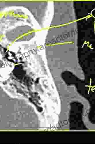
Temporal Bone CT and MRI Anatomy: A Comprehensive Atlas

The temporal bone, located at the base of the skull, is a captivating structure that houses crucial organs for hearing and balance. Its intricate anatomy poses challenges for medical professionals, making a comprehensive understanding of its CT and MRI anatomy essential for accurate diagnosis and effective treatment. This article serves as an in-depth guide, providing a detailed exploration of the temporal bone's anatomy, illuminated through high-resolution CT and MRI images.
The external anatomy of the temporal bone can be divided into several regions:
- Squamous part: Forms the posterolateral aspect of the temporal bone.
- Petrous part: Contains the inner ear structures.
- Mastoid process: Contains air cells that connect to the middle ear.
- Styloid process: A slender projection for muscle attachments.
- Jugular foramen: An opening for the passage of blood vessels and nerves.
The internal anatomy of the temporal bone is even more complex, including:
5 out of 5
| Language | : | English |
| File size | : | 6450 KB |
| Text-to-Speech | : | Enabled |
| Screen Reader | : | Supported |
| Enhanced typesetting | : | Enabled |
| Print length | : | 224 pages |
- Middle ear: Composed of the tympanic membrane, ossicles, and Eustachian tube.
- Inner ear: Houses the cochlea and vestibular system, responsible for hearing and balance.
- Facial nerve canal: A bony channel that houses the facial nerve.
- Carotid canal: A passageway for the carotid artery.
- Eustachian tube: A canal that connects the middle ear to the nasopharynx.
CT and MRI scans provide invaluable insights into the temporal bone's anatomy, allowing physicians to evaluate its structures in great detail.
CT scans utilize X-rays to create cross-sectional images of the bone. They excel in depicting bony structures, including the petrous part and mastoid air cells.
MRI scans employ magnetic fields and radio waves to generate images. They provide superior soft tissue contrast, making them ideal for visualizing the middle and inner ear structures, as well as the facial and vestibulocochlear nerves.
A thorough understanding of the temporal bone's anatomy is paramount in clinical practice. It facilitates the diagnosis and management of various conditions, such as:
- Otitis media: Infection of the middle ear.
- Mastoiditis: Infection of the mastoid air cells.
- Cholesteatoma: A skin growth in the middle ear.
- Facial nerve paralysis: Damage to the facial nerve.
- Acoustic neuroma: A benign tumor of the vestibular nerve.
Accurate imaging of the temporal bone guides surgical interventions, including:
- Tympanoplasty: Repair of the tympanic membrane.
- Mastoidectomy: Removal of infected mastoid air cells.
- Vestibular schwannoma resection: Surgical removal of an acoustic neuroma.
The temporal bone is an intricate structure that plays a vital role in hearing and balance. A comprehensive understanding of its CT and MRI anatomy is essential for medical practitioners involved in the diagnosis and treatment of temporal bone disFree Downloads. This guide provides a detailed exploration of the temporal bone's anatomy, empowering healthcare professionals to confidently navigate its complexities and optimize patient outcomes.
- Temporal Bone Imaging: A Comprehensive Atlas by F.H. Lingeman, W. Gullane, and T.C. Calcaterra.
- Otologic Imaging: A Textbook and Atlas by H.A. Harnsberger, H.W. Teixidó, and J.R. Dillon.
- The Temporal Bone: Anatomy, Radiology, Surgery by J.J. Fisch, R.A. Valvassori, and M.M. Mesolella.
5 out of 5
| Language | : | English |
| File size | : | 6450 KB |
| Text-to-Speech | : | Enabled |
| Screen Reader | : | Supported |
| Enhanced typesetting | : | Enabled |
| Print length | : | 224 pages |
Do you want to contribute by writing guest posts on this blog?
Please contact us and send us a resume of previous articles that you have written.
 Book
Book Novel
Novel Page
Page Chapter
 Text
Text Story
Story Genre
Genre Reader
 Library
 Paperback
 E-book
 Magazine
 Newspaper
 Paragraph
 Sentence
 Bookmark
 Shelf
Shelf Glossary
 Bibliography
 Foreword
 Preface
 Synopsis
 Annotation
 Footnote
 Manuscript
 Scroll
 Codex
Codex Tome
Tome Bestseller
 Classics
 Library card
 Narrative
 Biography
 Autobiography
 Memoir
 Reference
 Encyclopedia
 Michael Riera
 Gerald Posner
 David Tracy
 Alexander Bely
 Monika Pavlickova
 Nancy Dufresne
 Ian Peate
 Jackie Notman
 Matthew Mcclure
 Katherine Simpson
 Asad Bashey
 Nita Leland
 Eugene D Frank
 Charles Morgan
 W Leydhecker
 Ruth Forman
 Lita Epstein
 Dr Jagannath Dixit
 Marla Tetsuka
 Jennifer Cook
Light bulbAdvertise smarter! Our strategic ad space ensures maximum exposure. Reserve your spot today!

 Winston HayesConstructivist Developmental and Experiential Approaches: Uncover the Power...
 Cody BlairUnveiling the Synergies: German-Japanese Interchange of Data Analysis Results...
 Dwight BellDiscover the Intricacies of the Aging Auditory System with Springer Handbook...
 Edward BellFollow ·18.2k
 Dan BrownFollow ·19.3k
 Jeff FosterFollow ·3k
 Joseph ConradFollow ·12.2k
 Quincy WardFollow ·15.2k
 Edwin CoxFollow ·7.8k
 Jake CarterFollow ·15.1k
 Eric HayesFollow ·2.2k
 Ashton Reed
Unveiling the Silent Pandemic: Bacterial Infections and...
Bacterial infections represent...
 Brent Foster
Finally, Outcome Measurement Strategies Anyone Can...
In today's...
 Brett Simmons
Unlocking the Secrets to Entrepreneurial Excellence:...
Empowering...

 Eugene Powell
Our Search For Uncle Kev: An Unforgettable Journey...
Prepare to be captivated by...
5 out of 5
| Language | : | English |
| File size | : | 6450 KB |
| Text-to-Speech | : | Enabled |
| Screen Reader | : | Supported |
| Enhanced typesetting | : | Enabled |
| Print length | : | 224 pages |


